
Researchers create model of early human heart development from stem cells
Making it visible with ZEISS confocal microscope system
Researchers at the University of California, Berkeley, in collaboration with scientists at the Gladstone Institutes, have developed a template for growing beating cardiac tissue from stem cells, creating a system that could serve as a model for early heart development and a drug-screening tool to make pregnancies safer.*Confocal microscopy was performed with ZEISS LSM 710.
http://blogs.zeiss.com/microscopy/news/ ... -zeiss.mp4
Researchers at UC Berkeley and Gladstone Institutes have shown that human induced pluripotent stem cells can differentiate and self-organize into cardiac microchambers when spatially confined. The center (red) shows heart muscle cells while the perimeter (green ring) shows a type of connective tissue that attaches to the surface and holds the microchamber to the culture dish. (Video by Zhen Ma, UC Berkeley)
In experiments published on July 14, 2015 in the journal Nature Communications, the researchers used biochemical and biophysical cues to prompt stem cells to differentiate and self-organize into micron-scale cardiac tissue, including microchambers.
“We believe it is the first example illustrating the process of a developing human heart chamber in vitro,” said Kevin Healy, a UC Berkeley professor of bioengineering, who is co-senior author of the study with Dr. Bruce Conklin, a senior investigator at the Gladstone Institute of Cardiovascular Disease and a professor of medical genetics and cellular and molecular pharmacology at UC San Francisco. “This technology could help us quickly screen for drugs likely to generate cardiac birth defects, and guide decisions about which drugs are dangerous during pregnancy.”
Screening for drug toxicity
To test the potential of the system as a drug-screening tool, the researchers exposed the differentiating cells to thalidomide, a drug known to cause severe birth defects. They found that at normal therapeutic doses, the drug led to abnormal development of microchambers, including decreased size, problems with muscle contraction and lower beat rates compared with heart tissue that had not been exposed to thalidomide.
“We chose drug cardiac developmental toxicity screening to demonstrate a clinically relevant application of the cardiac microchambers,” said Conklin. “Each year, as many as 280,000 pregnant women are exposed to drugs with evidence of potential fetal risk. The most commonly reported birth defects involve the heart, and the potential for generating cardiac defects is of utmost concern in determining drug safety during pregnancy.”
The new milestone comes nearly four months after Healy and other UC Berkeley researchers publicly debuted a system of beating human heart cells on a chip that could be used to screen for drug toxicity. However, that heart-on-a-chip device used pre-differentiated cardiac cells to mimic adult-like tissue structure.
In this new study, the scientists mimicked human tissue formation by starting with stem cells genetically reprogrammed from adult skin tissue to form small chambers with beating human heart cells. Conklin’s lab at Gladstone, an independent, nonprofit life science research organization affiliated with UC San Francisco, supplied these human induced pluripotent stem cells for this study.
The undifferentiated stem cells were then placed onto a circular-patterned surface that served to physically regulate cell differentiation and growth.
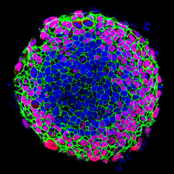
When grown on a geometrically confined surface, human induced pluripotent stem cells go through differentiation with distinct spatial patterning. The pluripotency biomarker (OCT4) is shown in red, the epithelial biomarker (E-cadherin) in green, and the nuclei in blue. This illustrates how biophysical cues determine stem cell fate and spatial patterning. (Image by Zhen Ma, UC Berkeley)
Location, location, location
By the end of two weeks, the cells that began on a two-dimensional surface environment started taking on a 3D structure as a pulsating microchamber. Moreover, the cells had self-organized based upon whether they were positioned along the perimeter or in the middle of the colony.
Compared with cells in the center, cells along the edge experienced greater mechanical stress and tension, and appeared more like fibroblasts, which form the collagen of connective tissue. The center cells, in contrast, developed into cardiac muscle cells. Such spatial organization was observed as soon as the differentiation started. Center cells lost the expression of octamer-binding*transcription factor*4 (OCT4) and epithelial cadherin (E-cadherin) faster than perimeter cells, which are critical to the development of heart tissue.
“This spatial differentiation happens in biology naturally, but we demonstrated this process in vitro,” said study lead author Zhen Ma, a UC Berkeley postdoctoral researcher in bioengineering. “The confined geometric pattern provided biochemical and biophysical cues that directed cardiac differentiation and the formation of a beating microchamber.”
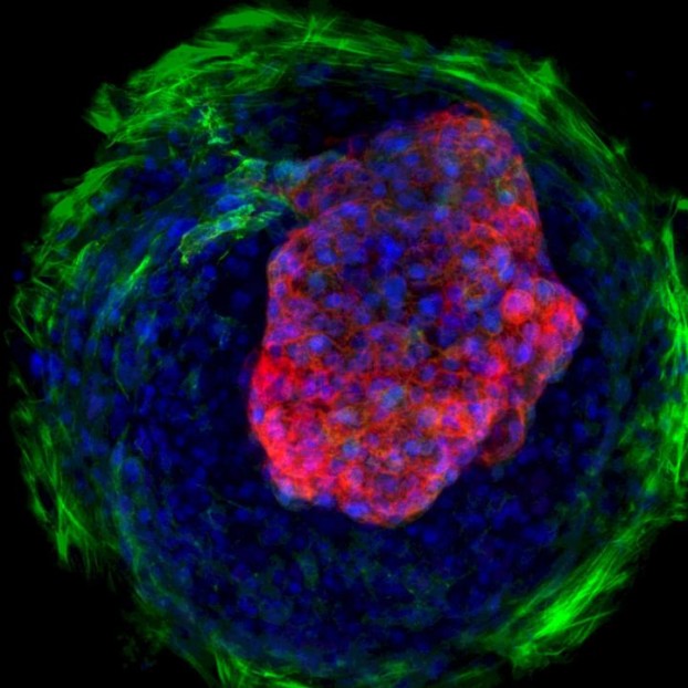
When spatially confined, differentiating human induced pluripotent stem cells self-organize into cardiac microchambers. Myofibroblasts are green, cardiomyocytes are red and the nuclei are blue. Cardiac microchambers can be used to study the toxicity of drugs that affect early human heart development.*(Image by Zhen Ma, UC Berkeley)Could eventually replace animal models
Modeling early heart development is difficult to achieve in a petri dish and tissue culture plates, the study authors said. This area of study has typically involved the dissection of animals at different stages of development to study the formation of organs, and how that process can go wrong.
“The fact that we used patient-derived human pluripotent stem cells in our work represents a sea change in the field,” said Healy. “Previous studies of cardiac microtissues primarily used harvested rat cardiomyocytes, which is an imperfect model for human disease.”
The researchers pointed out that while this study focused on heart tissue, there is great potential for use of this technology to study other organ development.
“Our focus here has been on early heart development, but the basic principles of patterning of human pluripotent stem cells, and subsequently differentiating them, can be readily expanded into a broad range of tissues for understanding embryogenesis and tissue morphogenesis,” said Healy.
The National Institutes of Health and a Siebel Postdoctoral Fellowship helped support this research.
RELATED INFORMATION
- Self-organizing human cardiac microchambers mediated by geometric confinement (study in Nature Communications)
- Bioengineers put human hearts on a chip to aid drug screening
- Confocal microscopes by ZEISS – Meet our new LSM 8 family!
About the Gladstone Institutes
To ensure our work does the greatest good, the Gladstone Institutes focus on conditions with profound medical, economic, and social impact—unsolved diseases of the brain, the heart, and the immune system. Affiliated with the University of California, San Francisco, Gladstone is an independent, nonprofit life science research organization that uses visionary science and technology to overcome disease.
About UC Berkeley
The University of California was chartered in 1868, and what would become its flagship campus was soon established at Berkeley. It was born out of a vision in the State Constitution of a university that would “contribute even more than California’s gold to the glory and happiness of advancing generations.” UC Berkeley claims 22 Nobel Laureates, seven of whom are current faculty members. The campus enrolls more than 36,000 undergraduate and graduate students, and has more than 1,500 full-time and 500 part-time faculty members in more than 130 academic departments and more than 110 interdisciplinary research units and field stations. Today, UC Berkeley is considered one of the nation’s most prestigious universities – public or private – and is internationally recognized for its distinguished record of world-class scholarship, innovation and concern for the betterment of our world.
More...